
Dermoscopy is known as surface microscopy, dermatoscopy or epiluminescence microscopy and it consists of an imaging method that allows increase the lesions 10X to 120X leading a visualization of some structures located within the skin (epidermis and dermis).
Thus, dermoscopy is useful for recognize skin tumors and the main indication is to diagnose cutaneous melanomas in early phases of evolution and infiltration. See Figure 1.
Generally, the accuracy for diagnosing melanoma with naked eyes is about 75 to 80% for dermatologists and even lower for residents and general practitioners. Using dermoscopy, this accuracy can reach 90% or more depending on the experience of the examiner.
Thus, the dermoscopy sometimes is not able to distinguish melanocytic nevi (moles) from melanomas since initial melanoma lesions may not present the usual malignant criteria for its diagnosis, they are called featureless melanomas or uncharacteristic melanomas.In this context, the Total Body Mapping and Digital Dermoscopy(TBMDD) have been developedfor the high-risk patient’s evaluation. This exam consists of photographs of the whole body combined with dermoscopy of melanocytic lesions. This methodology allowed the image storage and monitoring of the pigmented lesions during time for a very early melanoma diagnosis.
Based on the principle that the benign lesions do not modify, and melanomas undergo considerable changes over time (due to proliferation), the TBMDD revealed to be efficient to diagnose uncharacteristic melanomas.The initial photography serves of base for a better follow up and permits the detection and removal of only suspected new lesions or changed lesions therefore, reducing costs, avoiding unnecessary surgeries, and reassuring high-risk patients.
Currently, anew generation diagnostic tool, the Intelligent Total Body Scanner (iToBos) System is being manufactured as part of aninternational project for developing an artificial intelligence (AI) assisted diagnostic platform for the early detection of melanoma. This project involves 18 partners, who are all located within the European Union (including UK and Israel), except for The University of Queensland, located in Brisbane, Australia. And has received funding from the European Union’s Horizons 2020 Innovative funding program, under the Health, Demographic Change and Wellbeing section.This platform will include a total body scanner, powered with high-resolution cameras that allow clinical and dermoscopy images, and a Computer Aided Diagnostics (CAD) tool to combine data sources such as medical records, genomics data and imaging data. The iToBoS will be designed with the purpose of developing a new tool for supporting clinical decisions by integrating various diagnostic data for early detection of melanoma in high-risk patients.
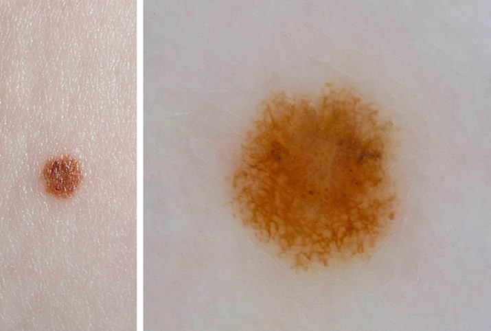
Figure 1. On the left, clinical image of an in situ melanoma. On the right, dermoscopy image (10X) of the same lesion.
G.Rezze, J.Malvehy
Hospital Clinic of Barcelona, IDIBAPS
References:
Thomas L, Puig S. Dermoscopy, Digital Dermoscopy and Other Diagnostic Tools in the Early Detection of Melanoma and Follow-up of High-risk Skin Cancer Patients. Acta DermVenereol. 2017;Suppl 218:14-21. doi: 10.2340/00015555-2719. Epub 2017 Jul 5. PMID: 28676882.
Salerni G, Carrera C, Lovatto L, Martí-Laborda RM, Isern G, Palou J, Alós L, Puig S, Malvehy J. Characterization of 1152 lesions excised over 10 years using total-body photography and digital dermatoscopy in the surveillance of patients at high risk for melanoma. J Am Acad Dermatol. 2012 Nov;67(5):836-45. doi: 10.1016/j.jaad.2012.01.028. Epub 2012 Apr 20.