
Most people who know about melanomas have a particular idea of what they look like: dark brown or black blotches or lumps. Many melanomas do look like this, so it’s what many machine learning algorithms are taught to look for.
But there are other, more challenging types of melanomas that the iToBoS clinical assistant will need to be trained on. One of these is amelanotic melanomas. “Amelanotic” means without melanin, the brown/black pigment that gives both moles and melanomas their colour. Instead, amelanotic melanomas are often pink or red in colour, and can easily be confused for other lesions like basal cell carcinomas or Bowen disease.
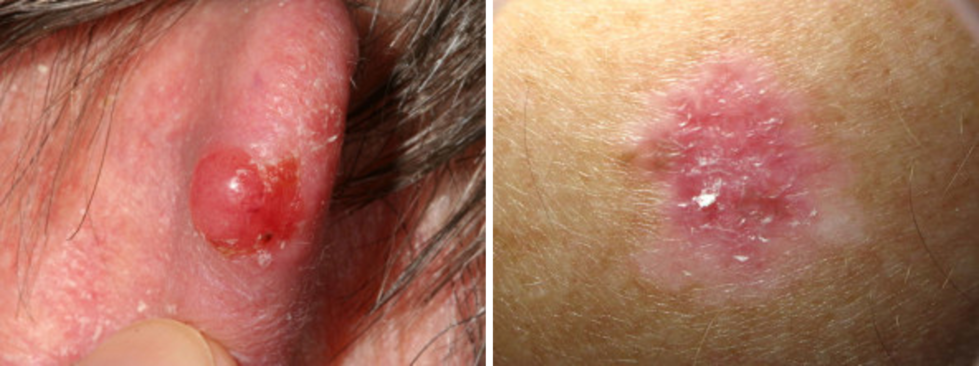
Image: amelanotic melanomas that could be mistaken for a basal cells’ carcinoma or Bowen disease. Image credit: dermnetnz.org (CC BY-NC-ND 3.0 NZ)
Often on closer inspection, there is some pigment in the lesion, but it seems “smudged” or out of focus, and only makes up a small part of the lesion. Often the only way to tell an amelanotic melanoma apart from other pale lesions is by closely looking at the blood vessels visible under dermoscopy. Amelanotic melanomas often have small blood vessels visible in a variety of shapes – coiled, hairpin-shaped, dots, and wavy lines, and areas of uniform, pinky-red.
However, patients themselves can provide a critical clue: if the lesion is new and growing fast. This is because many amelanotic melanomas are nodular in form and can “bulge out” from the skin suddenly.
Amelanotic melanomas have a similar prognosis to regular pigmented melanomas at a similar thickness, but because they are so easily missed, they are often not diagnosed until they are thick and ulcerated, leading to worse survival outcomes. Amelanotic make up 2-8% of all cutaneous melanomas, so it will be critical for iToBoS’s clinical assistant to learn spot these melanomas.
Katie Lee - University of Queensland.